Could a particle that slips through a person almost unnoticed reveal what lies inside? Muons routinely cross Earth’s atmosphere and pass through substantial amounts of matter. Scientists have used their paths to inspect volcanoes and hidden spaces in ancient structures. But soft tissue presents a harder riddle: it barely nudges a muon, and different tissues may nudge it by only slightly different amounts. Could those tiny deflections add up to a useful image—or would the result mostly reflect what the reconstruction software expected to find?
A muon’s passage is not perfectly straight. As it travels through matter, it undergoes many small interactions with atomic nuclei, scattering by an amount that depends on the material and how much of it the particle crosses. The imaging idea is to measure each muon’s track before and after an object, then use the changes in direction to infer a three-dimensional map of its scattering density. Dense materials create a strong signal. Soft tissue, made mostly of light elements and with modest differences in density, is a much quieter target.
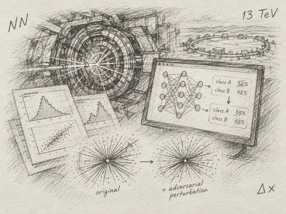
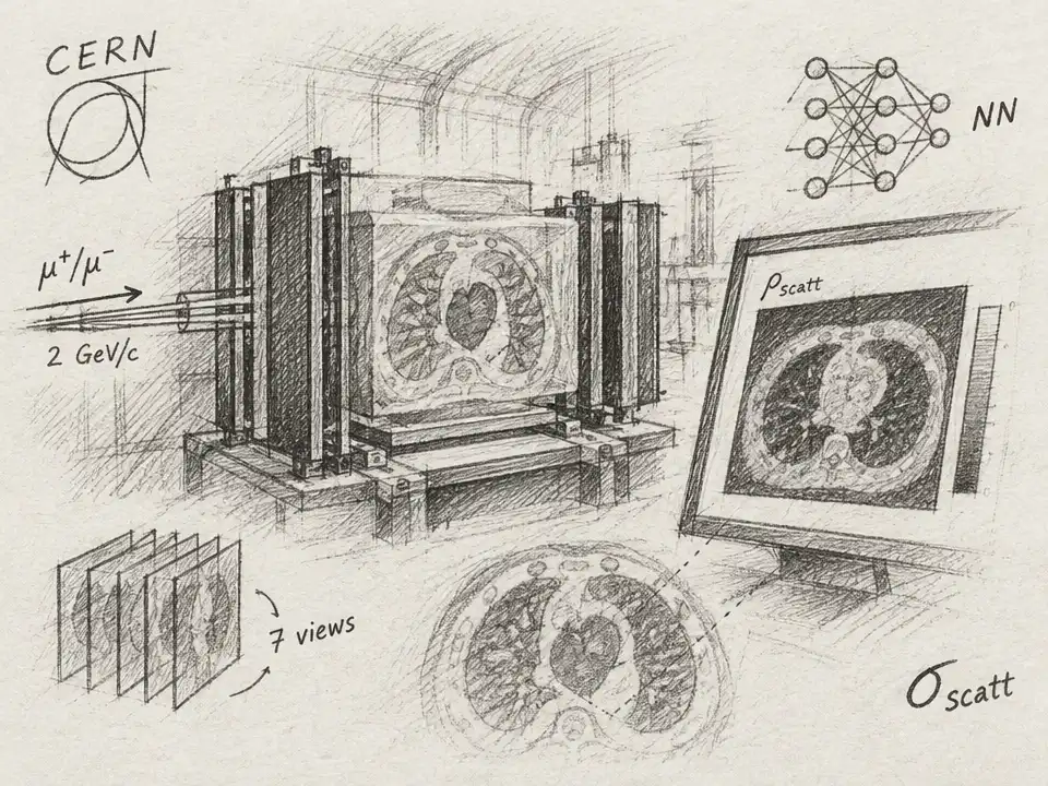
To test whether that quiet signal could be measured, researchers brought an anthropomorphic thorax phantom to the CERN Proton Synchrotron. The phantom contains materials designed to mimic tissue, lungs, blood, heart, and bone. A beam of positive muons with a momentum of 2 GeV/c passed through it. Two tracking detectors recorded each particle’s path before the phantom; two more recorded it afterward. The team turned the difference between those tracks into a scattering measurement, then rotated the phantom through seven projection angles.
The first clue was encouraging, but incomplete. From the scan, the researchers reconstructed a three-dimensional volume in which the tissue-equivalent region showed a consistent scattering signal. Yet the experiment did not see the whole phantom from every direction. The beam illuminated a narrow, structured patch, and the detectors had limited reach. Roughly 20% of the body was sampled from at least one angle. The region sampled from all seven—the part where the reconstruction could be quantitatively tested—was only about 0.8% of the phantom, a volume roughly 80 by 70 by 80 millimeters near the heart and mediastinum.
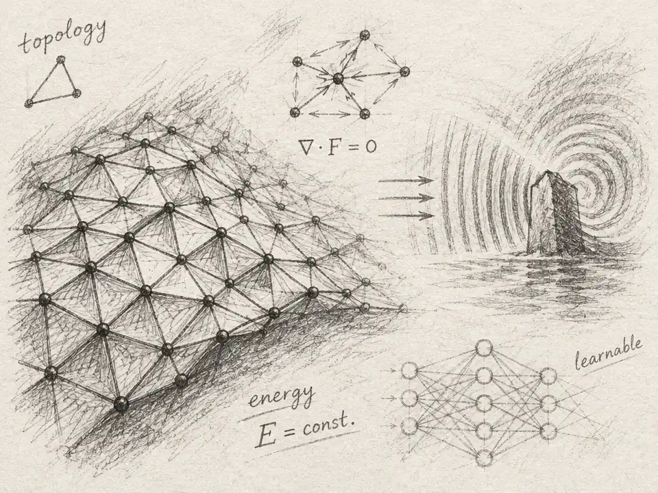
That limitation raises a rival explanation: perhaps the reconstruction did not extract a faint signal so much as smooth away the mess. The researchers first used a maximum-likelihood method to turn scattering measurements into a volume. That result was noisy and, with the restricted views, biased. They then passed it through a three-dimensional neural network trained exclusively on Monte Carlo simulations: computer-generated muon scans of randomized phantom geometries. A learned prior can use patterns across neighboring voxels to clean up noise, but it also makes it essential to ask whether the cleaned image is grounded in the actual beam data.
The team tested that concern in simulations designed to match the real detector acceptance and measured beam footprints. In this setting, the initial reconstruction overestimated the scattering density in the fully sampled core by 48%. After the network’s correction, the average came within 0.4% of the simulated ground truth across five held-out phantom geometries. Then came the key check: applying the network to the real scan without rescaling or recalibrating it produced a core average within 1% of the simulation-based expectation. Tightening the cut that removed unusually large scattering angles changed the network’s output by less than 1%, even though it shifted the raw reconstruction by 15%.
That agreement is evidence that the measured muons contain usable information about soft-tissue scattering. It is not proof that the method has already produced a clinical-quality picture. The quantitative test was in a largely uniform region, and the network’s reference scale came from simulations. Outside the fully sampled core, ribs, sternum, and spine appeared in roughly the right places, but sparse views and alignment uncertainties stretched some structures into streaks. The paper’s authors identify denser angular coverage and precise knowledge of the detector and patient positions as necessary steps toward reliable imaging of anatomical boundaries.
A smaller feature exposed the harder limit: statistics. The phantom included air-filled myocardial defects, but the reconstruction did not recover them. A muon passing through one such defect would travel about 27 millimeters through air instead of tissue along a total scattering path of roughly 250 millimeters, reducing its measured scattering by about 10%. In the fully sampled region, each one-millimeter column was crossed by an average of about 58 muons across the seven views. The resulting per-column uncertainty was about 13%—large enough to bury a signal of roughly 10%. The authors estimate that ten times as many muons could bring that defect signal to the edge of detectability.
The possible attraction is a small energy deposit. The researchers estimate that the scan delivered a local absorbed dose of about 2 microgray in the fully sampled region. They also estimate that a tenfold increase in statistics would raise that to about 20 microgray. Those figures are not directly comparable to clinical effective doses, which account for how radiation affects different organs; the paper stresses that a full dose assessment would need to include the beam’s uneven distribution and secondary radiation. And the test used an accelerator beam, not naturally occurring muons. Cosmic-ray muons are too scarce for practical medical imaging, while more compact, intense muon sources remain under development.
So the puzzle has a careful answer. Muons can carry enough information to measure soft-tissue scattering in a limited, well-sampled region, and the reconstruction’s match to the beam data survived useful checks. But seeing fine details means collecting more particles, controlling their paths, and proving that images of less uniform anatomy are trustworthy. This experiment did not deliver a new medical scanner. It showed that the weak signal is real—and mapped the obstacles between that clue and a usable image.