At the edge of a living cell, chemistry is happening on a scale too small for an ordinary camera to follow. Molecules shift, bonds break, and new substances form across the cell’s outer membrane. The action leaves clues in the way molecules vibrate—but those clues are faint. A new microscope technique makes them easier to see by turning each molecular vibration into a tiny, rapidly fading heat signal.
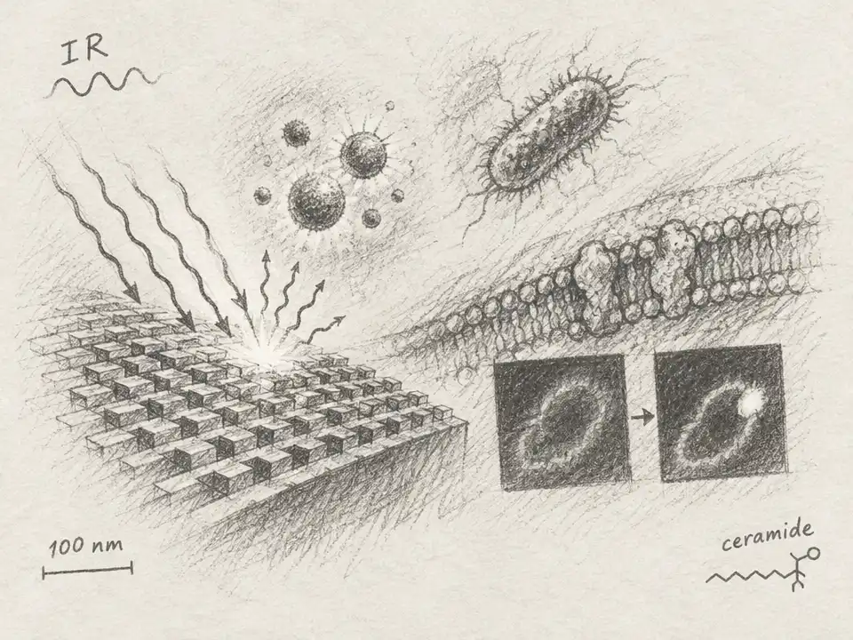
The method, called metasurface-driven mid-infrared photothermal imaging, or MetaMIP, begins with a useful fact: different chemical bonds absorb infrared light at characteristic frequencies. A molecule that absorbs the light warms slightly. Researchers can detect that temperature change with visible light, but in conventional methods the signal from small amounts of material can be hard to distinguish from background.
The team’s answer is a patterned gold surface on a transparent calcium fluoride base. Its nanoscale structures are designed to resonate with a chosen infrared frequency, concentrating the light close to the sample. Think of the surface as a tiny acoustic chamber, but for light: when its resonance matches a molecular vibration, more energy gathers where the target molecules sit. The pattern can be tuned for different chemical bonds.
That concentrated energy creates a brief thermal pulse. In the experiments, infrared light arrived in 500-nanosecond pulses, repeating 100,000 times per second. A visible 532-nanometer beam then read the sample’s response. In one version of the microscope, heating slightly changes how the sample scatters visible light; in another, it changes fluorescence. Either way, the visible signal tracks the infrared-absorbing molecules without relying on the visible beam to identify their chemistry.
The trick is not simply to make the sample hotter. It is to create a stronger temperature change and let it cool before the next pulse. Fluorescent probe beads next to the metasurface registered a peak rise of about 20 kelvin, compared with 5–7 kelvin on an unpatterned surface. Simulations predicted about 25 kelvin near the patterned structures and about 10 kelvin on the plain substrate. The measured heat relaxed with a characteristic time of about 6 microseconds; the pulse cycle left a 9.5-microsecond recovery interval.
That amplified response helped the researchers detect oleic acid at 100 nanomolar under their test conditions—about a 100-fold improvement over the micromolar sensitivity typically reported for conventional mid-infrared photothermal imaging. They also detected individual 100-nanometer plastic beads by their characteristic bond vibration. The measured image widths were 330 and 335 nanometers, so this is not a claim that the microscope resolves every 100-nanometer detail. It shows that objects that small can produce a detectable, chemically specific signal.
The team then tested individual Staphylococcus aureus bacteria. The cells were inactivated before imaging. When the infrared light targeted a vibration associated with proteins, the patterned surface produced clearer signals than conventional imaging. Just as important, changing the surface’s resonance changed the result: enhancement appeared when the resonance matched the targeted vibration, not when it was tuned to a different one. That control helped show the signal was tied to chemistry, rather than merely to the shape of the gold pattern.
The most revealing experiment took place at the surface of living HeLa cells. The researchers added an enzyme that breaks down sphingomyelin, a lipid in the cell membrane. The reaction leaves ceramide in the membrane and releases phosphocholine into the surrounding liquid. As the reaction proceeds, a phosphate-related infrared signal near 1085 inverse centimeters should weaken. A signal near 1030 inverse centimeters, associated with carbon–oxygen vibrations, could grow as ceramide forms.
That is broadly what the microscope recorded. Across cells imaged at 0, 10, and 20 minutes after the enzyme was added, the 1085 signal faded over time. In a closer comparison of the same region of an individual cell before and 10 minutes after treatment, the researchers also saw the 1030 signal increase. Its pattern became concentrated in the central region where the cell membrane contacted the metasurface, while the phosphate-associated signal declined more broadly. The authors describe the increase as consistent with ceramide formation—not as a uniquely definitive identification of ceramide.
That difference in spatial patterns is the hidden-world payoff. A measurement averaged across a whole cell could show that the chemical signals changed, but miss that they changed in different places. MetaMIP instead offers a way to map where bond-specific signals rise or fall as a reaction unfolds at a cell’s boundary.
The view still has limits. The enhanced field is strongest close to the patterned surface, so the signal depends not only on molecular composition but also on how closely the cell membrane sits against it. A change in contact or spacing can alter the measured intensity. The researchers say that more spectral bands and careful calibration would strengthen chemical assignments and quantitative measurements. Their demonstration also focuses on the basal membrane—the part nearest the substrate—not every reaction throughout the cell.
For now, MetaMIP is best understood as a new kind of close-up: not a complete movie of a cell, but a way to make faint molecular changes visible in a specific patch of its surface. By shaping infrared light and reading the heat that follows, the microscope turns a fleeting vibration into a map—and gives researchers a chance to watch chemistry take place where a living cell meets its world.