Inside a living cell, some of the most important chemical messages are fleeting. Nitric oxide and other reactive molecules can appear near a synapse or mitochondrion, then diffuse away. To understand what they do, a sensor needs to be close to the source—and quiet enough not to disturb it. A surprising possibility is to make the sensor out of a protein the cell can build and place for us, then use the protein’s own quantum spin to detect nearby chemistry.
Two recent arXiv studies sketch different parts of that idea. One builds a microscope designed to work with both diamond-based and biological spin probes in cell samples. The other calculates whether a fluorescent-protein spin sensor could detect neural radicals—and finds that the protein’s own molecular vibrations may overwhelm the signal. Together, they tell a crossover story, but not a claim that a working neural quantum sensor is already here.
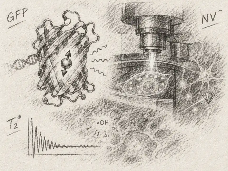
The biological sensor at the center of the second study is based on enhanced yellow fluorescent protein, or EYFP. Its light-absorbing chromophore can host a metastable spin-1 state: a quantum state with three spin sublevels that can be controlled with microwaves and read through light. Because the protein can be genetically encoded and attached to a chosen molecular target, it offers something a nanodiamond probe may not: a route to placing a sensor very close to a specific protein or radical-producing enzyme. The researchers describe a placement within about 3 nm of an enzyme’s molecular structure.
The proposed readout borrows from quantum sensing. A radical has an unpaired electron, whose fluctuating magnetic field affects a nearby sensor spin. Those fluctuations can speed up the sensor’s return toward its relaxed state. Measure that relaxation time, called T1, and—in principle—learn about the concentration of radicals nearby. The trick is to tell a chemical signal apart from the sensor’s own background relaxation.
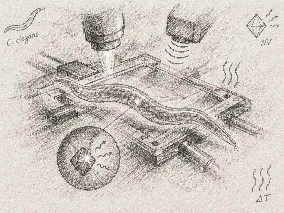
That is where the first study’s microscope enters the picture. Its authors built a confocal imaging and spin-control platform around a stationary cell sample, combining laser excitation, microwave control, magnetic-field delivery, and single-photon detection. They demonstrated spin-resonance measurements on a nitrogen-vacancy center in diamond, including a resonance near 2.87 GHz and a relaxation time of about 1.3 ms. They also imaged HeLa cells expressing MagLOV, a different magneto-sensitive fluorescent protein, and showed an image containing both MagLOV-expressing cells and nanodiamonds taken up by the cells.
There is an important boundary between that demonstration and the more ambitious vision. In the paper’s detailed account, the MagLOV experiments establish fluorescence imaging—not spin-resonance readout of the biological qubit in the cell. The authors describe biological-qubit spin measurements and entanglement between biological and diamond spins as future steps. And the EYFP sensor analyzed in the second paper is not the same protein as MagLOV. The studies are complementary pieces of a research direction, not one experiment that has already measured neural chemistry with an engineered protein qubit.
The second paper asks what might stop that next step. Its calculations include photon-counting noise, limits on how many photons a molecule can emit before bleaching, and the protein qubit’s intrinsic relaxation. At room temperature, the modeled sensor falls about six orders of magnitude short of sensitivity to micromolar bursts of nitric oxide, and about eight orders short of resting nanomolar levels. Those are theoretical detection limits, not measurements of a sensor in a living brain.
The culprit is vibration. The protein and its chromophore are not rigid; they vibrate, and those motions can disrupt the spin state. The authors estimate that a two-phonon Raman process dominates room-temperature relaxation over a direct process by about 720:1. Their model suggests that roughly doubling the effective stiffness of the chromophore’s surroundings could raise T1 to around 100 µs, a possible route toward micromolar sensing if the photon budget also improves. For nanomolar sensing, they identify a harder ceiling: suppressing that vibration-driven process alone cannot get past a modeled direct-process limit of 79 µs, while the required relaxation time is around 1 ms.
That distinction turns the calculation into a design brief. Genetic targeting may solve the problem of where the sensor sits, but it does not automatically solve how long its quantum state survives, how many useful photons it produces, or how quickly it can follow a changing signal. A sensor that integrates light for longer may detect weaker chemistry, but it can miss fast fluctuations. The paper’s estimates therefore point not to one magic improvement, but to a combination of a more stable molecular environment and better photon collection.
For now, the quantum microscope and the EYFP sensitivity model mark a frontier, not a finished instrument for neuroscience. Their crossover is still striking: cell biology offers a way to position a spin-bearing molecule, while quantum physics supplies a method for reading its response to nearby magnetic noise. The next test is whether engineers can make that molecular spin last long enough—and shine brightly enough—to report what a cell is doing before the signal disappears.